- Issues
- About the Journal
- News
- Cooperation
- Contact Info
Issue. Articles
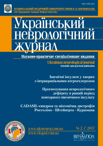
¹2(43) // 2017

1. Reviews
|
Notice: Undefined index: picture in /home/vitapol/ukrneuroj.vitapol.com.ua/en/svizhij_nomer.php on line 74 Notice: Undefined index: pict in /home/vitapol/ukrneuroj.vitapol.com.ua/en/svizhij_nomer.php on line 75 Experimental study on the correlation of lipid metabolism impairment and cerebral blood circulationN. S. TurchynaO. O. Bogomolets National Medical University, Kyiv |
|---|
Keywords: atherosclerosis, experiment.
Notice: Undefined variable: lang_long in /home/vitapol/ukrneuroj.vitapol.com.ua/en/svizhij_nomer.php on line 188
2. Reviews
|
Notice: Undefined index: picture in /home/vitapol/ukrneuroj.vitapol.com.ua/en/svizhij_nomer.php on line 74 Notice: Undefined index: pict in /home/vitapol/ukrneuroj.vitapol.com.ua/en/svizhij_nomer.php on line 75 Basic pathogenetic mechanisms of demyelination in the central nervous system and its correction limitationsL. D. Pichkur, S. A. Verbovs’ka, S. T. Akinola, G. E. ChitaevaSI «Institute of Neurosurgery named after acad. A. P. Romodanov of NAMS of Ukraine», Kyiv |
|---|
Keywords: multiple sclerosis, experimental autoimmune encephalomyelitis, mechanisms of pathogenesis, demyelination, remyelination, current approaches for treatment.
Notice: Undefined variable: lang_long in /home/vitapol/ukrneuroj.vitapol.com.ua/en/svizhij_nomer.php on line 188
3. Reviews
|
Notice: Undefined index: picture in /home/vitapol/ukrneuroj.vitapol.com.ua/en/svizhij_nomer.php on line 74 Notice: Undefined index: pict in /home/vitapol/ukrneuroj.vitapol.com.ua/en/svizhij_nomer.php on line 75 Prospects of implementation of harm reduction strategy in health care of contemporary UkraineV. V. KorolenkoO. O. Bogomolets National Medical University, Kyiv |
|---|
Keywords: epidemiology, public health, WHO, Ukraine, harm reduction.
Notice: Undefined variable: lang_long in /home/vitapol/ukrneuroj.vitapol.com.ua/en/svizhij_nomer.php on line 188
4. Original researches
|
Notice: Undefined index: picture in /home/vitapol/ukrneuroj.vitapol.com.ua/en/svizhij_nomer.php on line 74 Notice: Undefined index: pict in /home/vitapol/ukrneuroj.vitapol.com.ua/en/svizhij_nomer.php on line 75 The structurally and functional characteristic of ischaemic stroke in patients with intracranial atherosclerosisO. E. Dubenko 1, V. V. Lebedynets 2, P. V. Lebedynets 1, D. P. Kovalenko 3, T. I. Nesterenko 31 Kharkiv Medical Academy of Postgraduate Education |
|---|
Methods and subjects.The 451 patients with ischaemic stroke who had neuroimaging angiography of intracranial arteries were examined. We selected 51 with angiographic signs of intracranial atherosclerosis(male/female 35/16, mean age 64.1 ± 10.1). Cardiovascular risk factors and brain imaging were compared with 57 patients examination with extracranial carotid atherosclerotic stenosis > 50 % (male 34/female 23, mean age 68.8 ± 9.1).
Results. The predominant risk factors was arterial hypertension but its frequency did not differ in patients with intra-and extracranial atherosclerosis. Patients with intracranial atherosclerosis more often suffered from diabetes mellitus — 72.5 % than patients with extracranial carotid atherosclerotic stenosis — 28.0 % (p < 0.01), myocardial infarction was observed in 39.2 % and 14.0 % (p < 0.001) respectively, ischaemic stroke history was in 45.0 % and 10.5 % (p < 0.05). Atherogenic dyslipidemia was more specific for patients with extracranial atherosclerosis. Predominant localization of intracranial atherosclerosis was middle cerebral artery — in 49.0 %, intracranial internal carotid — in 29.4 %, basilar artery in 13.7 % and intracranial part of the vertebral artery — in 9.8 % patients. According to the brain imaging, large territorial infarction involving two lobes occurs twice more often in stroke due to intracranial atherosclerosis (15.6 % and 7.0 %, p = 0.05). The severity of white matter hyperintensities (Fazekas grade ≥ 2) was more intensive in stroke patients with intracranial atherosclerosis — 37.2 % and 19.3 % (p < 0.05).
Conclusions. The risk factors profile and neurovisualisation pattern are significantly different in stroke patients with intracranial or extracrania latherosclerosis. It is important for treatment and prevention of recurrent stroke.
Keywords: intracranial atherosclerosis, ischaemic stroke, risk factors.
Notice: Undefined variable: lang_long in /home/vitapol/ukrneuroj.vitapol.com.ua/en/svizhij_nomer.php on line 188
5. Original researches
|
Notice: Undefined index: picture in /home/vitapol/ukrneuroj.vitapol.com.ua/en/svizhij_nomer.php on line 74 Notice: Undefined index: pict in /home/vitapol/ukrneuroj.vitapol.com.ua/en/svizhij_nomer.php on line 75 Clinical-paraclinical and neuropsychological features among patients with recurrent cerebral ischemic hemispheric strokeO. A. Kozyolkin, L. V. NovikovaZaporizhzhya State Medical University |
|---|
Methods and subjects. The study involved 49 patients (22 men and 27 women) aged from 44 to 85 years in the acute period of re-CIHS. The first clinical observation group included 26 patients with re-CIHS subcortical localization, and the second group consisted of 23 patients with re-CIHS of cortical-subcortical localization. The study of the dynamics of neurological deficit was assessed with the NIHSS (National Institutes of Health Stroke Scale) on 1st—3rd and 10th—13th days and the clinical and social outcome of the acute period of re-CIHS was assessed by modified Rankin scale (on the 21st day of the disease). The neuropsychological study included the verification of cognitive impairment using the Mini-Mental State Examination (MMSE), the Montreal Cognitive Assessment (MoCA) and the Frontal Assessment Battery (FAB) scales. All patients underwent computed tomography in order to verify the localization of focus of cerebral ischemia regarding cortical structures, and to detailed morphological changes. The following computer tomography parameters were taken into account: the location, the size of the focus of cerebral ischemia, the expansion of the ventricular system, the indices and sizes of the third and fourth ventricles, the dislocation processes, the type of hydrocephalus, the localization of the cyst, the presence and severity of leukoaraiosis, signs of cerebral cortex atrophy.
Results. Patients with subcortical localization of re-CIHS in the right hemisphere were characterized more severe neurological deficit and worse medical and social outcome of the acute period of the disease. The cognitive deficit among patients with re-CIHS cortical-subcortical localization was significantly more pronounced by the MMSE, MoCA and FAB scales during all observation periods compared with patients who had subcortical re-CIHS. A strong correlation was found between parameters of computed tomography, the severity of the stroke and the level of cognitive deficits.
Conclusions. It was found that patients with re-CIHS subcortical localization had a more pronounced neurological deficit and less favorable medical and social outcome of the acute period of the disease. Cognitive deficit, under the remised stroke of cortical- subcortical localization, was more severe according to MMSE, MoCA and FAB scales during all observation periods. There is a significant correlation of CT data, stroke severity stage and cognitive deficit level.
Keywords: recurrent cerebral ischemic hemispheric stroke, subcortical localization, cortical-subcortical localization, cognitive impairments.
Notice: Undefined variable: lang_long in /home/vitapol/ukrneuroj.vitapol.com.ua/en/svizhij_nomer.php on line 188
6. Original researches
|
Notice: Undefined index: picture in /home/vitapol/ukrneuroj.vitapol.com.ua/en/svizhij_nomer.php on line 74 Notice: Undefined index: pict in /home/vitapol/ukrneuroj.vitapol.com.ua/en/svizhij_nomer.php on line 75 Neurohormonal criteria for neurological deficit level prediction in early recovery period of cerebral ischemic hemispheric strokeS. O. MedvedkovaZaporizhzhya State Medical University |
|---|
Methods and subjects. Complex examination was performed for 77 patients (mean age of patients was 57.9 ± 0.9 years) in early recovery period of CHIS using National Institute of Health Stroke Scale, Barthel Index, modified Rankin Scale on the 10th, 30th, 90th and 180th day of disease, CT scan. The determination of melatonin serum level and plasma concentration of serotonin was on the 10th, 30th days of the disease. The melatonin/serotonin ratio (MSR) = melatonin serum level/plasma concentration of serotonin was calculated.
Results. The most informative parameters for prediction of the value by means of NIHSS ≥ 5 score on the 90th day of CIHS are: dynamics of serotonin plasma level on the 30th day (AUC = 0.82, p < 0.05), the level of MSR on the 30th day (AUC = 0.77, p < 0.05), serotonin plasma level on the 30th day (AUC = 0.70, p < 0.05) and on the 10th day (AUC = 0.65, p < 0.05); for prediction the level of neurological deficit by means of NIHSS ≥ 5 score on the 180th day of CIHS — the dynamics of MSR on the 30th day (AUC = 0.73, p < 0.05), level of MSR on the 30th day (AUC = 0.72, p < 0.05), dynamics of serotonin plasma level on the 30th day (AUC = 0.70, p < 0.05) and its level on the 10th day (AUC = 0.65, p < 0.05).
Conclusions. Serotonin plasma level on the 30th day ≤ 0.18 mcmol/l is the predictor of value according to NIHSS ≥ 5 score on the 90th day of disease (AUC = 0.72, p < 0.05; sensitivity = 66.7 %, specificity = 72.1 %); dynamics of serotonin plasma level ≤ –0.262 on the 30th day of CIHS is the predictor of neurological deficit as for NIHSS ≥ 5 score on the 90th day of disease (AUC = 0.70, p < 0.05; sensitivity = 66.7 %, specificity = 72.1 %) and on the 180th day of disease (AUC = 0.70, p < 0.05; sensitivity = 66.7 %, specificity = 81.4 %).
Keywords: ischemic hemispheric stroke, serotonin, melatonin, prognosis.
Notice: Undefined variable: lang_long in /home/vitapol/ukrneuroj.vitapol.com.ua/en/svizhij_nomer.php on line 188
7. Original researches
|
Notice: Undefined index: picture in /home/vitapol/ukrneuroj.vitapol.com.ua/en/svizhij_nomer.php on line 74 Notice: Undefined index: pict in /home/vitapol/ukrneuroj.vitapol.com.ua/en/svizhij_nomer.php on line 75 Risk factors of the spastic forms of the infantile cerebral palsy depending on the infant age gestationV. V. Abramenko 1, O. E. Kovalenko 21 Ukrainian Medical Rehabilitation Center for Children with Organic Diseases of the Nervous System of Health Ministry of Ukraine, Kyiv |
|---|
Methods and subjects. The article presents the results of the clinical examination of 193 families with children with spastic forms of cerebral palsy to study the risk factors of the pathology origination. Families were separated into two groups according to the gestational age of a new born child: one group (families with cerebral palsy in full-term children) and control group (families with cerebral palsy in pre-term children). The groups were compared according to the family, obstetric history of the mother and pre- and post-natal data, and the presence of bad habits from their parents.
Results. It was defined that the presence and combination of such factors as parents’ age (over 30 years), mother’s complicated obstetric history (previous labours, miscarriages, abortions) and complicated labours allow to predict pre term baby birth with a risk of cerebral palsy development. Insufficient attention towards infants’ health and underestimation of neurological deficiency, especially in pre-term babies, prevented in time treatment and rehabilitation which caused cerebral palsy development.
Conclusions. Parents’ age over 30 years, mother’s complicated obstetric history and complicated labours allow to predict baby birth with a risk of cerebral palsy development. In case of full term babies, insufficient attention towards infants’ health state assessment and neurological deficiency were observed.
Keywords: cerebral palsy, spastic form, risk factor, central nervous system, full-term, pre-term.
Notice: Undefined variable: lang_long in /home/vitapol/ukrneuroj.vitapol.com.ua/en/svizhij_nomer.php on line 188
8. Original researches
|
Notice: Undefined index: picture in /home/vitapol/ukrneuroj.vitapol.com.ua/en/svizhij_nomer.php on line 74 Notice: Undefined index: pict in /home/vitapol/ukrneuroj.vitapol.com.ua/en/svizhij_nomer.php on line 75 Quality of children’s life with cerebellar medulloblastoma after combined treatmentV. V. Morgun 1, L. M. Verbova 1, L. L. Marushchenko 1, A. V. Shaversky 1, M. O. Marushchenko 21 SI «Institute of Neurosurgery named after acad. A. P. Romodanov of NAMS of Ukraine», Kyiv |
|---|
Methods and subjects. The results of 297 children treatment with cerebellum of different age groups were analyzed. The age of children ranged from 3 months up to 18 years (M = 7.6 ± 2.1 years). 33 (11.1 %) patients were children of 0 — 3 years old, 97 (32.6 %) — of 3 — 7 years, 114 (38.4 %) children — of 7 — 11 years, 53 (17.8 %) children — of 12 — 18 years. All patients underwent pre- and postoperative clinical and instrumental examination, including CT, cerebral MRI and spinal cord, and studies of lumbar cerebrospinal fluid according to the recommendations. In the early postoperative period 31 (10.8 %) patients died. The catamnesis from 1 month to 10 years was followed in 266 (84.6 %) patients. Radiation therapy (RT) was performed in 186 (66.9 %) children, chemotherapy (CT) — in 121 (45.4 %). Functional status was assessed according to the Karnovsky (Lansky) scale and Orlov Yu. A. scale.
Results. At hospitalization in 48 % of patients the Karnovsky index was 60 — 70 points or higher, in 45 % — 50 — 60 points, in 7 % of cases 30 — 40 points. In more than 80 % of cases, the size of the MB was more than 3 cm in diameter, predominantly involving the IVth ventricle and spreading to the adjacent structures of the brain stem. Total tumor removal was performed in 113 (40.6 %) cases, subtotal in 149 (53.6 %), partial or tumor biopsy in 18 (5.8 %). In 49 (16.5 %) patients, postoperative complications were revealed. In the younger age group metastasis was observed in 18.0 % of observations, in the elder groups an average data was 13.8 %.
Conclusions. The median survival (MS) did not exceed 12 — 24 months in all ages after surgical treatment of the cerebellum MB in children. After CT or RT this index was 18 — 24 months and with full implementation of the protocols of adjuvant treatment in children of 4 — 18 years of age the MS was 36 months. In children of the first 3 years the MS was 12 months and 18 months respectively.
Keywords: cerebellum medulloblastoma, children, combined treatment, median survival, quality of life.
Notice: Undefined variable: lang_long in /home/vitapol/ukrneuroj.vitapol.com.ua/en/svizhij_nomer.php on line 188
9. Original researches
|
Notice: Undefined index: picture in /home/vitapol/ukrneuroj.vitapol.com.ua/en/svizhij_nomer.php on line 74 Notice: Undefined index: pict in /home/vitapol/ukrneuroj.vitapol.com.ua/en/svizhij_nomer.php on line 75 Does electroencephalographic study help to differentiate organic and functional changes in patients with the effects of traumatic brain injury?L. L. Chebotariova, O. S. SolonovichSI «Institute of Neurosurgery named after acad. A. P. Romodanov of NAMS of Ukraine», Kyiv |
|---|
Methods and subjects. The study was conducted on 70 patients with mild concussion or contusion aged 18 to 45 years. The comparison group included 40 healthy people of the same age. Clinical, neurological, neuropsychological testing scales, digital electroencephalography, the cognitive evoked potentials were applied.
Results. Patients with mild traumatic brain injury in the intermediate and late period had dysfunction in following cognitive domains: attention, memory, delayed recall, symptoms of anxiety and depression. Neurophysiological features of bioelectrical activity of the cerebral cortex included increasing in peak P300 latency in 42.86 % patients; reducing the amplitude of cognitive evoked potentials — 45.71 %; tendency to disruption of major cortical rhythms, spatial inversion alpha rhythm, its frequency and amplitude. Method of binary logistic regression allowed to determine predictors of cognitive impairment in patients with mild traumatic brain injury, based on the results of neuropsychological, neurophysiological research.
Conclusions. Digital EEG and cognitive evoked potentials methods should be performed using as the diagnostic screening in patients with mild traumatic brain injury in the intermediate and late periods. As predictors of cognitive impairment, a set of statistically significant deviations from the normative values of neurophysiological parameters was proposed: increased latency and decreased amplitude of cognitive evoked potentials, changes in the electroencephalogram. It is well known that digital EEG and the cognitive evoked potentials methods have the great sensitivity and accuracy, but their specificity in diagnostic of traumatic disorders requires further research.
Keywords: mild traumatic brain injury, cognitive impairment, diagnostics, cognitive evoked potentials.
Notice: Undefined variable: lang_long in /home/vitapol/ukrneuroj.vitapol.com.ua/en/svizhij_nomer.php on line 188
10. Experimental researches
|
Notice: Undefined index: picture in /home/vitapol/ukrneuroj.vitapol.com.ua/en/svizhij_nomer.php on line 74 Notice: Undefined index: pict in /home/vitapol/ukrneuroj.vitapol.com.ua/en/svizhij_nomer.php on line 75 Efficiency weld the damaged peripheral nerve rat according to estimates sciatic nerve functional indexV. I. Tsymbaliuk 1, 2, V. Yu. Molotkovets 1, 2, V. V. Medvediev 2, B. M. Luzan 2, T. I. Petryv 11 SI «Institute of Neurosurgery named after acad. A. P. Romodanov of NAMS of Ukraine», Kyiv |
|---|
Methods and subjects. Experimental animals were CD-1 albino rats-male (350 — 450 g, 7 months); trauma was the section of the left sciatic nerve in the middle third. Examined experimental groups were: 1 (neurotomy, n = 6), 2 (neurotomy + epineural neurorrhaphy, n = 15), 3 (neurotomy + epineural welding, n = 18). Method of research is the definition of a functional index sciatic nerve at 1, 3 and 5 months after the injury.
Results. High-frequency electric welding of epineurum provides reliable connection of nerve stumps. The approach efficiency, in terms of remote monitoring of the sciatic nerve functional index, is comparable with epineural neurorrhaphy. Significant regeneration of paretic limb motor function, in the case of a welded connection epineural sciatic nerve stumps, accounts for the first 3 months of posttraumatic period when epineural neurorrhaphy — 3 — 5th month.
Conclusions. Epineural welded connection sciatic nerve stumps is accompanied by faster recovery of motor function paretic limb. According to overall productivity, it is not inferior to epineural neurorrhaphy.
Keywords: neurotomy, neurorrhaphy, weld biological tissues, functional index sciatic nerve, regeneration of peripheral nerves.
Notice: Undefined variable: lang_long in /home/vitapol/ukrneuroj.vitapol.com.ua/en/svizhij_nomer.php on line 188
11. Original researches
|
Notice: Undefined index: picture in /home/vitapol/ukrneuroj.vitapol.com.ua/en/svizhij_nomer.php on line 74 Notice: Undefined index: pict in /home/vitapol/ukrneuroj.vitapol.com.ua/en/svizhij_nomer.php on line 75 Diagnostics of cerebral autosomal dominant arteriopathy with subcortical infarcts and leukoencephalopathy (CADASIL syndrome) in combination with Rossolimo — Curschmann — Steinert myotonic dystrophyI. A. Grygorova 1, K. A. Leshchenko 1, Yu. V. Severyn 2, I. V. But 11 Kharkiv National Medical University |
|---|
Keywords: CADASIL syndrome, myotonic dystrophy, diagnostics.
Notice: Undefined variable: lang_long in /home/vitapol/ukrneuroj.vitapol.com.ua/en/svizhij_nomer.php on line 188
12. TO HELP PRACTICING PHYSICIANS
|
Notice: Undefined index: picture in /home/vitapol/ukrneuroj.vitapol.com.ua/en/svizhij_nomer.php on line 74 Notice: Undefined index: pict in /home/vitapol/ukrneuroj.vitapol.com.ua/en/svizhij_nomer.php on line 75 Clinical approaches to interpretation of research results vertebrogenic myelopathy at different levels of the spinal cordZ. I. Zavodnova, M. G. MatiushkoO. O. Bogomolets National Medical University, Kyiv |
|---|
Methods and subjects. The study involved 54 patients with VM: 35 (64.8 %) men, 19 (35.2 %) women. Patients are divided into groups depending on the level of the spinal cord lesion: cervical segment — 20 patients (44.4 %), thoracic — 12 patients (22.2 %), lumbar — 18 (33.4 %). All patients underwent a complete clinical examination, including MRI of the spinal cord, electroneuromyography (ENMG), etc.
Results. Discogenic myelopathy was diagnosed in 50 patients, 4 patients had discogenic radiculomyelopathy. The pain severity was pronounced in the lesion of two parts simultaneously in the lumbar spinal cord, moderately pronounced was in the chest, which is confirmed by the scale JOA (p < 0.05). The treatment was effective for VM in the cervical section: 43.7 % of the patients noted improvement (p < 0.05). The use of the ENMG method in the diagnosis of VM could be recommended as an opportunity to confirm the diagnosis.
Conclusions. Based on clinical and instrumental studies conducted in patients with VM at different levels of the spinal cord, it is revealed that the lumbar and cervical spinal cord are more often affected. In the same departments more pronounced pain syndrome is observed. Conservative treatment of VM is currently not effective, as 18.3 % of patients with lumbar spine and 25.5 % — with simultaneous lesion of the two departments were discharged unchanged.
Keywords: vertebrogenic myelopathy, levels of spinal cord lesion, clinical manifestations, electroneuro-myography.
Notice: Undefined variable: lang_long in /home/vitapol/ukrneuroj.vitapol.com.ua/en/svizhij_nomer.php on line 188
Current Issue Highlights
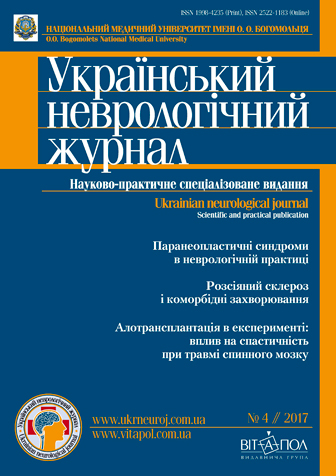
¹4(45) // 2017

Paraneoplastic syndromes in neurological practice
E. G. Dubenko, L. I. Kovalenko
Analysis of comorbid diseases in patients with multiple sclerosis
Ò. ². Nehrych, Ê. Ì. Hychka
Comparative analysis of the rat’s paretic limb spasticity against the background of spinal cord injury, adult olfactory bulb and fetal cerebellum tissue allotransplantation
V. I. Tsymbaliuk 1, 2, V. V. Medvediev 2, Yu. Yu. Senchyk 3, N. G. Draguntsova 1
Log In
Notice: Undefined variable: err in /home/vitapol/ukrneuroj.vitapol.com.ua/blocks/news.php on line 50

